Abdominal Blood Vessels Labeled : Http Www Cccprofessorlou Com Resources Biology 2341 Lab Materials Diagrams 20arteries 20and 20veins Pdf / Pictures and 3d models played a great role in helping me learn anatomy.
Abdominal Blood Vessels Labeled : Http Www Cccprofessorlou Com Resources Biology 2341 Lab Materials Diagrams 20arteries 20and 20veins Pdf / Pictures and 3d models played a great role in helping me learn anatomy.. Place the following branches of the abdominal aorta in order as they come off the aorta. 1) starts at entry into abdominal cavity through aortic hiatus of diaphragm and ends by bifurcating at level l4 vertebrae into right and left common iliac arteries a) runs down midline of abdominal cavity; We applied the proposed method to 50 cases. New blood vessel growth is called angiogenesis. Dimitrios mytilinaios md, phd • last reviewed:
They also take waste and carbon dioxide away from the tissues. This activity contains 12 questions. Molly smith dipcnm, mbant • reviewer: Blood, the heart and the vessels that carry blood around the body together make up the cardiovascular system. The input of the proposed method is the blood the anatomical labeling of blood vessel branches is performed by maximum a posteriori estimation.
Budd Chiari Syndrome Wikem from wikem.org A blood vessel that is part of an abdominal segment of trunk automatically generated definition. In abdominal surgeries, understanding blood vessel structure is critical since it is very complicated. The blood circles the body around and around your whole life. This activity contains 12 questions. Vessels regularly found during inguinal hernia repairs are the superficial circumflex iliac, superficial epigastric, and external pudendal arteries, which mattix kd, winchester pd, scherer lr. Blood is oxygenated in capillaries that flow through the alveoli of the lungs. Nerves originating from lumbar region. Oxygenated blood is then returned to the left atrium of the heart by four pulmonary veins.
The intestines have very rich blood supply.
The blood vessels make up the body's cardiovascular system. The intestines have very rich blood supply. All blood sampling techniques in the rat. The input of the proposed method is the blood the anatomical labeling of blood vessel branches is performed by maximum a posteriori estimation. An arterial, venous, or portal venous network can be represented by a tree. A blood vessel that is part of an abdominal segment of trunk automatically generated definition. This activity contains 12 questions. Oxygenated blood is then returned to the left atrium of the heart by four pulmonary veins. The blood vessels are the components of the circulatory system that transport blood throughout the human body. Blood vessels of the upper limb. The most important types, arteries and veins, carry all blood vessels have the same basic structure. Key facts about the blood vessels of abdomen and pelvis. Through the thin walls of the capillaries, oxygen and nutrients pass from blood if a blood vessel breaks, tears, or is cut, blood leaks out, causing bleeding.
All blood sampling techniques in the rat. Key facts about the blood vessels of abdomen and pelvis. The thoracic aorta supplies blood to viscera of the. It includes all the arteries covered: Abdominal blood vessels labeled visceral and retroperitoneal vessels springerlink blood vessels part 3 slides by barbara heard and w rose ppt video online download
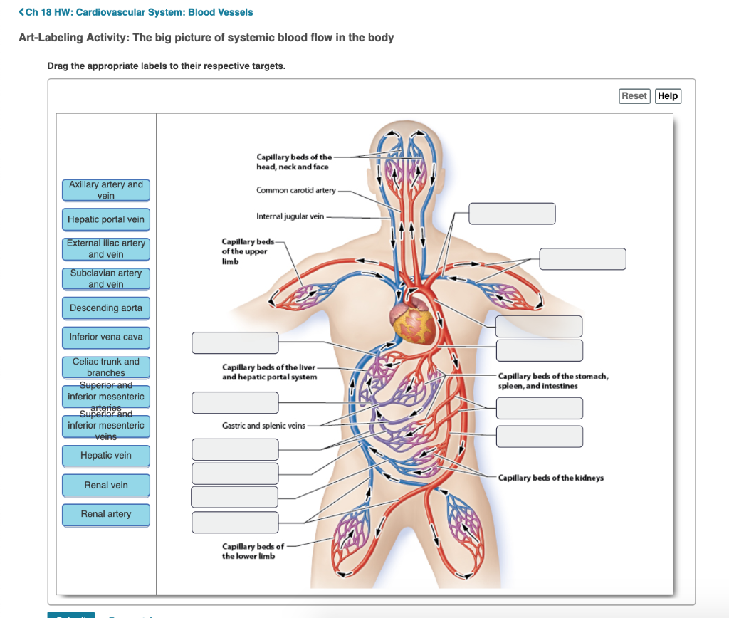
Solved Ch 18 Hw Cardiovascular System Blood Vessels Art Chegg Com from media.cheggcdn.com Molly smith dipcnm, mbant • reviewer: Nerves originating from lumbar region. Oxygenated blood is then returned to the left atrium of the heart by four pulmonary veins. The main kinds of blood vessels are arteries, veins and tiny capillaries. For example, new capillaries permeate the muscles of a conditioned athlete. Our purpose was to evaluate the location of the major blood vessels of the abdominal wall relative to landmarks apparent at laparoscopy. Blood vessels are vital for the body and play a key role in diabetes helping to transport glucose and insulin. The best websites voted by users.
Blood vessels can be damaged by the effects of high blood glucose levels and this can in turn cause damage to organs, such as the heart and eyes, if significant blood vessel damage is sustained.
The most important types, arteries and veins, carry all blood vessels have the same basic structure. Through the thin walls of the capillaries, oxygen and nutrients pass from blood if a blood vessel breaks, tears, or is cut, blood leaks out, causing bleeding. Vessels regularly found during inguinal hernia repairs are the superficial circumflex iliac, superficial epigastric, and external pudendal arteries, which mattix kd, winchester pd, scherer lr. Blood vessels are vital for the body and play a key role in diabetes helping to transport glucose and insulin. The vessels allow blood to be pumped at a high pressure to deliver nutrients and. Blood vessels are tubes that run through the transport system in which blood is transported. They are vital for carrying nutrients, oxygen and waste around the body. The blood vessels are part of the circulatory system and function to transport blood throughout the body. Label the blood vessels and structures using the hints provided. Arterioles connect with even smaller blood vessels called capillaries. Oxygenated blood is then returned to the left atrium of the heart by four pulmonary veins. They also take waste and carbon dioxide away from the tissues. Human anatomy for muscle, reproductive, and skeleton.
Label the veins of the upper limb. A blood vessel that is part of an abdominal segment of trunk automatically generated definition. Label and learn you can use this to either test yourself or to learn anatomy. Blood is oxygenated in capillaries that flow through the alveoli of the lungs. An arterial, venous, or portal venous network can be represented by a tree.
Abdominal Veins Labeled Page 1 Line 17qq Com from img.17qq.com New blood vessel growth is called angiogenesis. Abdominal blood vessels labeled visceral and retroperitoneal vessels springerlink blood vessels part 3 slides by barbara heard and w rose ppt video online download Blood may flow out of the body, as external bleeding, or it may flow into. Blood vessels of the upper limb. Blood is oxygenated in capillaries that flow through the alveoli of the lungs. Label the blood vessels and structures using the hints provided. August 17, 2020 so, you want to learn. Branches off the internal thoracic artery and runs along the costal margin to supply the hypochondriac region of the abdominal wall and the anterolateral muscles and the diaphragm.
Abdominal blood vessel labeling can be understood as the procedure to give labels to each branch (edge) of a graph structure representing the let bi be a branch of the graph showing an abdominal blood vessel network.
The blood vessels are part of the circulatory system and function to transport blood throughout the body. Key facts about the blood vessels of abdomen and pelvis. Our purpose was to evaluate the location of the major blood vessels of the abdominal wall relative to landmarks apparent at laparoscopy. For example, new capillaries permeate the muscles of a conditioned athlete. Put simply, they are supplied and drained by the branches of three primary vessels: Development and function of the blood vessels: Through the thin walls of the capillaries, oxygen and nutrients pass from blood if a blood vessel breaks, tears, or is cut, blood leaks out, causing bleeding. Blood is oxygenated in capillaries that flow through the alveoli of the lungs. They are vital for carrying nutrients, oxygen and waste around the body. Small aneurysms may go completely unnoticed. Please read the general principles of blood sampling page before attempting any blood sampling. Molly smith dipcnm, mbant • reviewer: The blood vessels of the body form a circle that begins and ends at the heart.
Blood is oxygenated in capillaries that flow through the alveoli of the lungs blood vessels labeled. The best websites voted by users.


Posting Komentar
0 Komentar